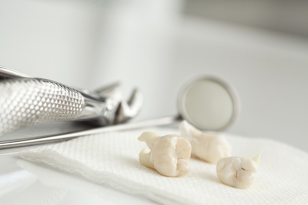
Prior research has documented associations between tooth loss and cognitive decline. Building on this previous work, a recent npj Aging study assessed the specific brain regions that are impacted by tooth loss and the associated causes.
Background
As the global aging population is increasing, age-related dementia and cognitive decline prevalence are increasing rapidly. Age-related tooth loss is also common, which influences dietary intake. Recent research has highlighted a link between oral health, diet, and cognitive abilities, which motivates further research in this area to understand the complex interplay between them better.
Several studies have highlighted the association between tooth loss and cognitive decline. However, it is still unexplained which specific brain regions are affected by tooth loss and what the potential underlying mechanisms are.
More research is needed to understand the impact of tooth loss on dietary patterns in cognitively normal persons and to what extent this channel explains cognitive decline and brain atrophy.
About the study
Addressing the aforementioned gap in the literature, the present study presents evidence from Japan on the links between oral functionality (use of dentures and tooth loss), dietary consumption, decline in cognitive ability, and dementia. The association between tooth loss and brain volume differences, in the case of dementia and mild cognitive impairment (MCI), was examined. To understand the potential role of diet in cognitive decline, the changes in dietary patterns post-tooth loss in cognitively normal individuals were studied.
A comprehensive and unique approach was adopted in this study, whereby detailed dietary assessments, dental examinations, general cognitive analysis, and magnetic resonance imaging (MRI) analysis were combined. The elderly Japanese cohort comprised 919 participants (510 women and 409 men) with an average age of 71.5 years.
Among the study participants, 17.7% belonged to the MCI group, and 2.6% constituted the dementia group. Heterogeneities across cognitive impairment statuses with respect to sex, age, duration of formal education, and prevalence of diabetes mellitus were studied.
Study findings
No significant association was noted between tooth loss and cognitive impairment, irrespective of denture use. However, the hippocampal volume was significantly reduced, and the white matter hypointensity (WMH) was significantly increased in the dementia and MCI groups relative to the normal control group.
The regional brain volumes of the lateral orbitofrontal cortex, insula, and posterior cingulate cortex were markedly reduced in the MCI group. For the dementia group, the parahippocampal gyrus, entorhinal cortex, and inferior temporal gyrus were significantly reduced.
Cognitively normal individuals who had less than ten teeth showed markedly smaller volumes of the superior parietal cortex, parahippocampal gyrus, middle temporal gyrus, bankssts, and lingual cortex. In such individuals, a bigger WMH volume was also noted relative to individuals with most residual teeth. It is important to note that greater WMH volume and atrophy of the parahippocampal gyrus, observed in individuals with tooth loss, are both characteristics of dementia.
The periodontal ligaments in natural teeth could be driving these results. These ligaments are connected to the trigeminal nerve that transmits sensory information to the brain. Loss of the periodontal ligaments could, therefore, lead to lower brain volume, and dentures are not able to make up for this deficit.
The parahippocampal gyrus and its neighboring structure, i.e., the hippocampus, could also be involved in the underlying mechanism. Delightful eating experiences can create and preserve episodic memories. With fewer teeth, an individual is less able to appreciate diverse flavors in food, making eating a less pleasurable experience.
This could have implications for vivid episodic memories, which, over time, could lead to reduced stimulation of the parahippocampal gyrus, subsequently leading to its atrophy.
Another finding was that tooth loss was associated with a reduction in the consumption of plant-based foods and an increase in the intake of fatty, processed foods. This could have contributed to cognitive decline and higher WMH volume through mechanisms such as inflammation, vascular dysfunction, and oxidative stress.
The association was strongest among individuals with fewer than ten teeth, which underscores the importance of maintaining at least this number of teeth for preserving both brain health and nutritional status as we age.
Conclusions
In sum, this study documented that tooth loss may be closely linked to changes in dietary patterns and brain atrophy, even in normal individuals. These changes could lead to dementia and further cognitive decline in the future. Therefore, proper management of oral health and consumption of a balanced diet could prevent neuropathological shifts associated with Alzheimer’s Disease.
The inability to establish causality between tooth loss and cognitive decline is a key limitation of this study. Additionally, the findings may not be readily generalizable to other populations as the study cohort comprises only elderly Japanese individuals.
The results could also have been influenced by confounders, such as the use of dental prosthetics, periodontal disease, and so on. Furthermore, recall bias could not be ruled out because information on diet was obtained through a self-administered diet history questionnaire.


