The cranial bone in the human body performs very important functions, such as protecting the brain and enabling the passage of the cranial nerves that are essential to physiological functioning. Critical-sized cranial defects can disrupt both the physical and psychological well-being of patients. Restoration of critical-sized cranial defects by cranioplasty is challenging for reconstructive surgeons, who prefer to use autologous bone grafts. The acquisition of autologous bone requires additional surgeries concomitant with risks such as free flap loss, infection, deep venous thrombosis, and nerve injury. These limitations necessitate the development of alternatives to autologous bone grafts for cranial defect restoration.
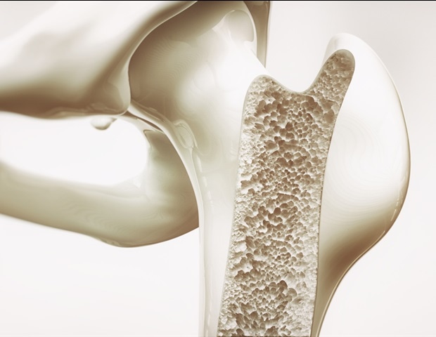
Biomaterials mimicking the composition and microstructure of natural bone are widely acknowledged to be ideal for bone defect regeneration The cranial bones are mainly composed of calcium phosphate, and they are typical flat bones, which are generally thin and broad with a flattened or curved surface. The flat bone has two outer compact tables made of cortical bone. The region between the two tables is called the diploe, which is composed of cancellous bone. Cortical bone has low porosity (5% to 10%) with interconnected tube-like pores, which are called Haversian canals and Volkmann canals. The cancellous bone is composed of irregular sponge-like trabeculae with a high specific surface area, and the mean surface curvature of the trabeculae surface is close to zero. The architectural characteristics of cancellous bone are similar to those of gyroid-type triply periodic minimal surface (TMPS) pore topology.
Inspired by the composition and structural features of cranial bones, scientists from South China University and Technology developed two flat-bone-mimetic β-tricalcium phosphate bioceramic scaffolds (Gyr-Comp and Gyr-Tub) by high-precision vat photopolymerization-based 3D printing. Both scaffolds had two outer layers and an inner layer with gyroid pores mimicking the diploe structure. The outer layers of Gyr-Comp scaffolds simulated the low porosity of outer tables, while those of Gyr-Tub scaffolds mimicked the tubular pore structure in the tables of flat bones. The Gyr-Comp and Gyr-Tub scaffolds possessed higher compressive strength and noticeably promoted in vitro cell proliferation, osteogenic differentiation and angiogenic activities compared with conventional scaffolds with cross-hatch structures. After implantation into rabbit cranial defects for 12 weeks, Gyr-Tub achieved the best repairing effects by accelerating the generation of bone tissues and blood vessels. The Gyr-Tub scaffolds have high prospects for treating cranial bone defects in clinical applications. This work provides an advanced strategy to prepare biomimetic biomaterials that fit the structural and functional needs of efficacious bone regeneration.
Source:
Journal reference:
Zhang, Y., He, F., Zhang, Q., Lu, H., Yan, S., & Shi, X. (2023). 3D-printed flat-bone-mimetic bioceramic scaffolds for cranial restoration. Research. doi.org/10.34133/research.0255.


